
Equine PET Scanner Making Big Strides at Santa Anita Park
This article originally appeared at vetmed.ucdavis.edu.
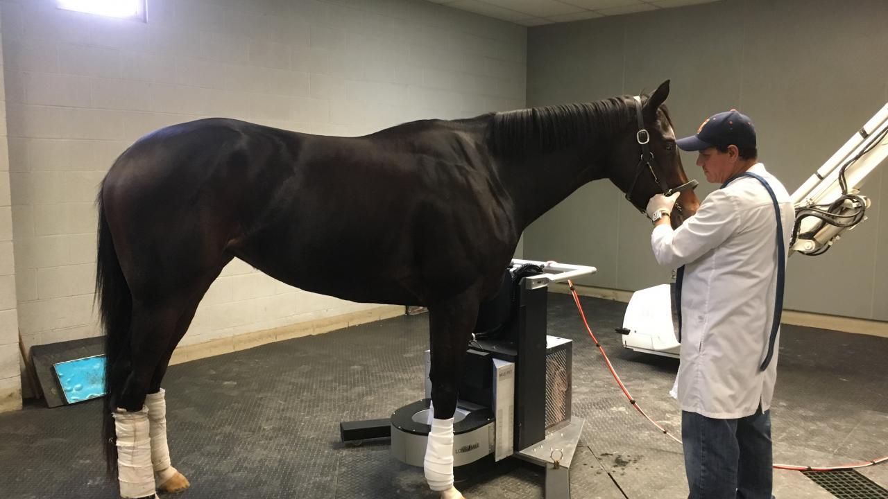
The equine Positron Emission Tomography (PET) scanner pioneered by the UC Davis School of Veterinary Medicine, in collaboration with LONGMILE Veterinary Imaging, is now in heavy use at Santa Anita Park in Southern California. In just over six months since the installation in December 2019, with the financial support from the Stronach Group, more than 100 scans have been performed with the “MILEPET” (Molecular Imaging of Limbs in Equids), the PET scanner specifically designed to acquire images on horses without the need to lay them down.
Overall, 65 different horses were imaged with the scanner, with several horses imaged multiple times, several weeks apart. In total, images of 186 front ankles, 28 hind ankles, 16 knees, and 11 feet were acquired. As shown by these numbers, the front ankles are the main area of interest, as these are the joints most commonly involved in severe injuries in racehorses.
The first objective after installation of the scanner at the racetrack was to complete a study, funded by the Grayson Jockey Club Research Foundation, aiming at assessing the value of PET for identification and follow-up of ankle injuries. The study called for 24 horses with signs of fetlock injuries to be imaged three times with the PET scanner, with follow-up scans six and 12 weeks after the initial scans. All 24 horses have now been enrolled, and over half of these horses have completed all their three scans.
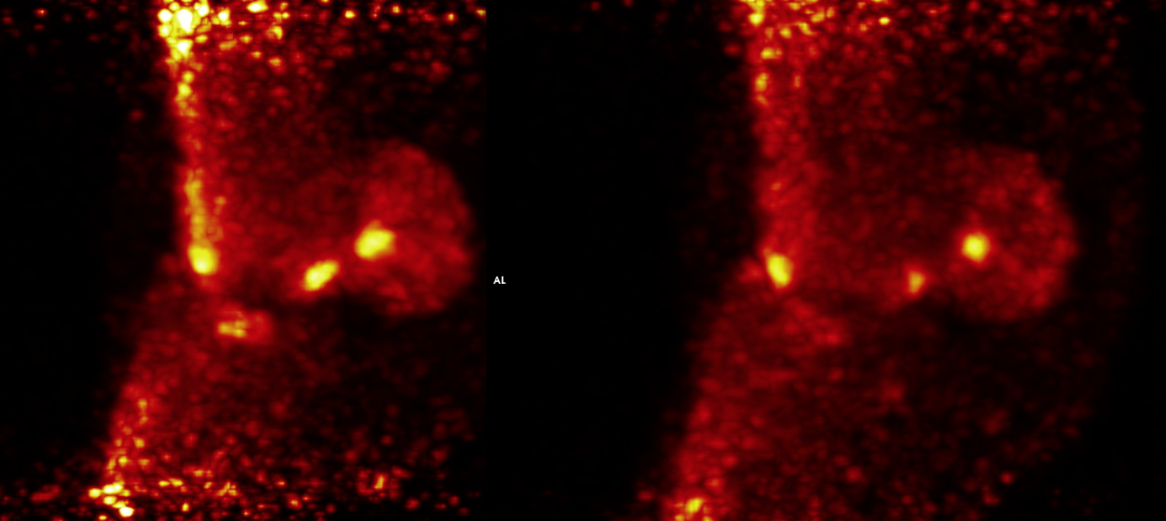
3D video of a PET scan of a front ankle showing multiple injuries in several bones. The yellow areas indicate precisely the sites of injury.
All scans were successful, providing very interesting preliminary results.
“The PET scans provide very precise information about the injury,” said Dr. Mathieu Spriet, associate professor of diagnostic imaging at UC Davis and lead veterinarian on the study. “With a classic ‘bone scan,’ we usually can associate the injury with one of the bones of the ankle, but with the PET, we can really localize the abnormality to a specific area in a bone.”
This is of particular importance, as injuries to specific areas of the ankle bones are known to predispose horses to catastrophic breakdown.
Another benefit of the PET scan is the ability to see changes over time.
“As we have repeated scans six weeks apart, we have seen marked improvement on many of the injuries following treatment and rest, whereas other injuries remained active,” continued Dr. Spriet. “Being able to assess the response of an injury is very helpful to decide the course of action: deciding whether a horse needs more time off, a different treatment, can go back to the track or should be retired from racing – these are very challenging decisions to make for the veterinarians at the track. The PET images have provided objective data to support these decisions.”
In addition to the 24 horses in the Grayson Jockey Club study, more than 40 horses have been scanned at the request of their veterinarians. Horses were sent in by 14 different veterinarians coming from 26 different trainers, demonstrating how broadly the technique has already been embraced by the Santa Anita racing community.
As a Magnetic Resonance Imaging (MRI) scanner was also installed at Santa Anita Park earlier this year, another on-going study, supported by the Oak Tree Foundation, aims at comparing PET and MRI findings in the ankles of racehorses. Twenty horses have been enrolled in this study with preliminary results demonstrating the complementarity of both techniques to provide the best assessment of various injuries.
Another study, supported by the Dolly Green Research Foundation, has recently started. This study aims at monitoring horses who are resuming training after being laid-up due to ankle injury. The goal is to assess the ankle on a regular basis to be able to adapt the training, if early signs of injury were to recur.
“It is very impressive how much has been accomplished and how much we have learned in the last six months, thanks to the support of key partners and the hard work of the technical staff and the veterinarians at Santa Anita Park,” said Dr. Spriet. “We have been able to establish this new technology as a reliable, high-precision diagnostic tool for racehorses.”
It is likely that the knowledge about PET will keep growing quickly. In addition to the different on-going studies at Santa Anita Park, this technology is now available outside of California, as a MILEPET scanner has just been installed at New Bolton Center, the equine veterinary hospital of the University of Pennsylvania. There are also on-going efforts to secure the funding to install another MILEPET at the UC Davis Veterinary Medical Center, where it would be available for racehorses from Golden Gate Fields and for sport horses across Northern California.